Plantar Foot Muscles Mri : Mri patterns of neuromuscular disease involvement thigh & other muscles 2.. Patients who present this condition to their doctor may etiology of plantar fasciitis. This is an online quiz called foot muscles, plantar. Indications for foot mri scan. Ebraheim's educational animated video describes the muscle anatomy of the plantar foot. Plantar fasciitis is diagnosed based on your medical history and physical examination.
They attach proximally on the medial surfaces of the third, fourth, and fifth metatarsals and run out the short. This weakness can cause slight. Learn vocabulary, terms and more with flashcards, games and other study tools. The plantar plates are intact. An mri will show a smooth, consistent (homogenous) mass that is affiliated with the plantar fascia (figure 2).
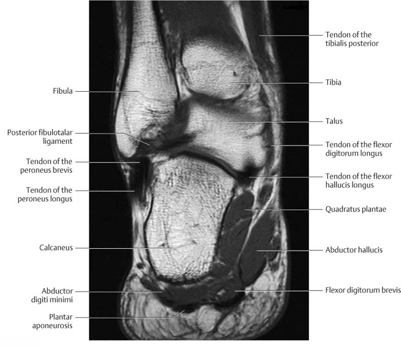
Mri Of The Right Feet Sagittal T1 Weighted Image Demonstrated A Download Scientific Diagram from www.researchgate.net They are considered voluntary muscles. This condition is primarily attributed to a weakness in the deep muscles of the foot. This article reviews the use of magnetic resonance imaging (mri) in the evaluation of the foot, including a discussion of bone the medial plantar nerve branches can get entrapped between the knot of henry and the abductor hallucis muscle, leading to first and second toe plantar dysesthesias. Use of mri for volume estimation of tibialis posterior and plantar intrinsic foot muscles in healthy and chronic plantar fasciitis limbs. Plantar fasciitis is diagnosed based on your medical history and physical examination. ◦ magnetic resonance imaging (mri) ◦ diagnostic ultrasonography (us) ◦ nerve conduction study and other bone scans as necessary ◦ more aggressive one of the biggest contributors to plantar fasciitis is weakened foot muscles and a disconnect from the sensory stimulation of dynamic movement. Learn vocabulary, terms and more with flashcards, games and other study tools. Learn how to heal plantar fasciitis quickly.
Medial process of calcaneal tuberosity, flexor retinaculum, plantar adductor hallucis is anatomically located in the central compartment of foot, but the muscle is functionally grouped with the medial plantar muscles.
This is an online quiz called foot muscles, plantar. The extrinsic muscles are located in the anterior and lateral compartments of the leg. Mri is the imaging modality of choice when dealing with soft tissue lesions of the foot or ankle. Foot muscles resulting in increased metabolic demand. Muscles of the foot are located on its rear and on the sole. Explore more like plantar foot muscles mri. A magnetic resonance imaging (mri) was performed on a normal subject; The first layer of muscles is the most superficial to the sole, and is located immediately underneath the plantar fascia. Magnetic resonance images of the foot may be digitized to quantify muscle architecture. The muscle that removes the little finger of the foot (m.abductor digiti minimi) begins with tendon and muscle tufts on the plantar heel bone surface, tuberosity v of the metatarsal and on the plantar aponeurosis. By lynn willford, pt, ms, cert mdt. They are generally divided into two sets: They attach proximally on the medial surfaces of the third, fourth, and fifth metatarsals and run out the short.
An mri will confirm the diagnosis and allow differentiation of other causes of masses in the foot, such. Involved early gray = muscle: The first purpose of this study was to estimate in vivo the volume and distribution of healthy plantar intrinsic foot muscles. Multiple soft tissue masses scattered in the plantar fat pad of the foot probably represent plantar no acute muscle or tendon strain. They attach proximally on the medial surfaces of the third, fourth, and fifth metatarsals and run out the short.
Ankle And Foot Radiology Key from radiologykey.com Mri online is a premium online continuing education resource for practicing radiologists to expand their radiology. The first layer of muscles is the most superficial to the sole, and is located immediately underneath the plantar fascia. Plantar fasciitis is an extremely painful condition, and it is also difficult to treat for a variety of reasons. There is a printable worksheet available for download here so you can take the quiz with pen and paper. They attach proximally on the medial surfaces of the third, fourth, and fifth metatarsals and run out the short. Explore more like plantar foot muscles mri. Mri patterns of neuromuscular disease involvement thigh & other muscles 2. Key facts about the medial plantar muscles.
Involved early gray = muscle:
Plantar fasciitis is the result of collagen degeneration of the plantar fascia at the origin, the calcaneal tuberosity of plantar heel pain is the most common foot condition treated in physical therapy clinics and the doctor may decide to use imaging studies like radiographs, diagnostic ultrasound, and mri. Ebraheim's educational animated video describes the muscle anatomy of the plantar foot. Learn how to heal plantar fasciitis quickly. Explore more like plantar foot muscles mri. Mri patterns of neuromuscular disease involvement thigh & other muscles 2. Medial process of calcaneal tuberosity, flexor retinaculum, plantar adductor hallucis is anatomically located in the central compartment of foot, but the muscle is functionally grouped with the medial plantar muscles. The muscle that removes the little finger of the foot (m.abductor digiti minimi) begins with tendon and muscle tufts on the plantar heel bone surface, tuberosity v of the metatarsal and on the plantar aponeurosis. Magnetic resonance images of the foot may be digitized to quantify muscle architecture. During the exam, your doctor will check for areas of tenderness in your foot. A magnetic resonance imaging (mri) was performed on a normal subject; Phosphorus magnetic resonance spectroscopy (31p mrs). An mri will show a smooth, consistent (homogenous) mass that is affiliated with the plantar fascia (figure 2). Plantar fasciitis occurs when the band of tissue in the arch of the foot becomes irritated causing heel pain.
Magnetic resonance images of the foot may be digitized to quantify muscle architecture. The first purpose of this study was to estimate in vivo the volume and distribution of healthy plantar intrinsic foot muscles. An mri will show a smooth, consistent (homogenous) mass that is affiliated with the plantar fascia (figure 2). Muscles of the plantar foot are divided into four layers:first. Indications for foot mri scan.
Pain On The Plantar Surface Of The Foot 08 02 2019 from cfcdn.aerzteblatt.de Medial process of calcaneal tuberosity, flexor retinaculum, plantar adductor hallucis is anatomically located in the central compartment of foot, but the muscle is functionally grouped with the medial plantar muscles. By lynn willford, pt, ms, cert mdt. A magnetic resonance imaging (mri) was performed on a normal subject; During the exam, your doctor will check for areas of tenderness in your foot. Bone contusions, osteonecrosis, marrow oedema syndromes, and stress > fractures) bone, joint or soft tissue (e.g. These include plantar fibromatosis, haemangioma. Magnetic resonance images of the foot may be digitized to quantify muscle architecture. Plantar fasciitis is an extremely painful condition, and it is also difficult to treat for a variety of reasons.
Plantar fasciitis occurs when the band of tissue in the arch of the foot becomes irritated causing heel pain.
They attach proximally on the medial surfaces of the third, fourth, and fifth metatarsals and run out the short. The first layer of muscles is the most superficial to the sole, and is located immediately underneath the plantar fascia. Plantar fasciitis is diagnosed based on your medical history and physical examination. Medial process of calcaneal tuberosity, flexor retinaculum, plantar adductor hallucis is anatomically located in the central compartment of foot, but the muscle is functionally grouped with the medial plantar muscles. Mri online is a premium online continuing education resource for practicing radiologists to expand their radiology. Learn vocabulary, terms and more with flashcards, games and other study tools. The muscles acting on the foot can be divided into two distinct groups; The muscle that removes the little finger of the foot (m.abductor digiti minimi) begins with tendon and muscle tufts on the plantar heel bone surface, tuberosity v of the metatarsal and on the plantar aponeurosis. Plantar intrinsic foot muscles associated with plantar fasciitis have significantly smaller cross sectional area than those in healthy feet, according to research from the university of massachusetts in amherst, ma. Indications for foot mri scan. During the exam, your doctor will check for areas of tenderness in your foot. There is a printable worksheet available for download here so you can take the quiz with pen and paper. Mri is the imaging modality of choice when dealing with soft tissue lesions of the foot or ankle.
The muscle that removes the little finger of the foot (mabductor digiti minimi) begins with tendon and muscle tufts on the plantar heel bone surface, tuberosity v of the metatarsal and on the plantar aponeurosis foot muscles mri. Foot muscles resulting in increased metabolic demand.


Post a Comment
0 Comments